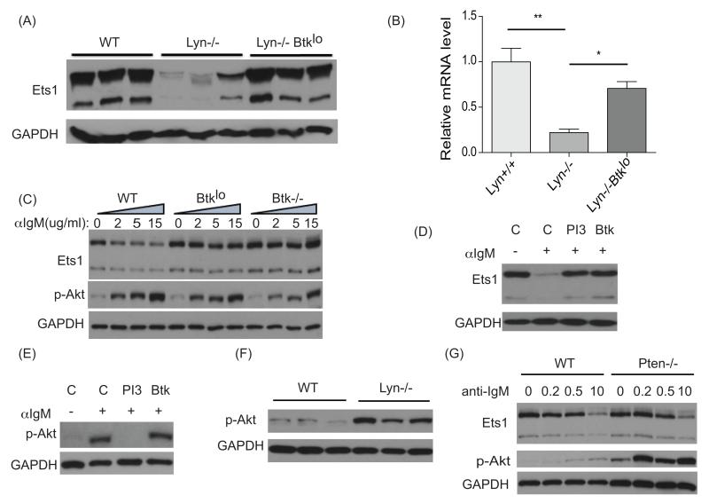
Figure 4. Ets1 downregulation in Lyn−/− B cells is dependent on signaling via Btk.
(A) Western blot for Ets1 and GAPDH with B cell lysates from wild-type (WT), Lyn−/− or Lyn−/−Btklo B cell mice (shown is a representative example of 3 independent experiments, n=4-5 mice of each genotype analyzed in total) (B) RT-qPCR analysis of Ets1 mRNA in B cells from wild-type, Lyn−/− or Lyn−/−Btklo mice (n=3 for each genotype). (C) Purified splenic B cells from wild-type (WT), Btklo or Btk−/− mice were rested at 37°C for 30 min before treatment with the indicated dose (μg/ml) of anti-IgM for 3.5 hours. Western blot for Ets1, p-Akt and GAPDH levels was performed with cell lysates. One of two similar experiments is shown. (D) Wild-type splenic B cells were rested at 37°C and pretreated with either DMSO vehicle control (labeled C), 5 μg/ml of a PI3K inhibitor Ly294002 (labeled PI3), 100 ng/ml of a Btk inhibitor PCI32765 (labeled Btk) for 1 hour, then stimulated with 10 μg/ml anti-IgM for 3.5 hours. Cell lysates were analyzed for Ets1 and GAPDH levels by Western blot. One of two similar experiments is shown. (E) To test the specificity of the inhibitors used in part D, cells were stimulated for 5 minutes and levels of phospho-S473 of Akt (activated Akt) were measured. One of two similar experiments is shown. (F) Western blot with lysates from wild-type (WT) or Lyn−/− B cells to show constitutive activation of the Akt pathway in the absence of Lyn. One of three similar experiments is shown. (G) Splenic B cells from wild-type (WT) or Pten−/− mice were rested at 37°C for 30 minutes followed by treatment with the indicated dose (μg/ml) of anti-IgM for 3.5 hours. Cell lysates were analyzed by Western blot. Shown is the data from one of two similar experiments. *p< 0.05, **p<0.01.

