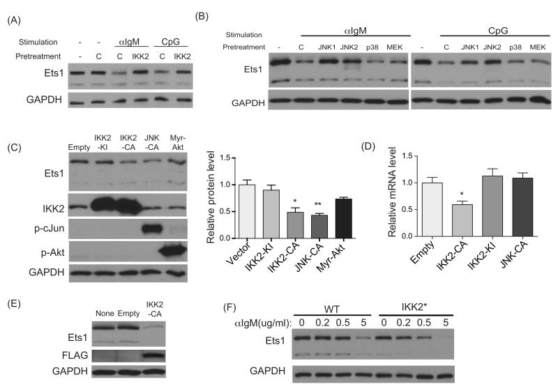
Figure 5. IKK2 and JNK both contribute to downregulating Ets1.
For experiments shown in parts A-B, wild-type splenic B cells were rested at 37°C and then pretreated with either DMSO vehicle control (labeled C) or with the inhibitors indicated below for 1 hour. Cells were then stimulated for 3.5 hours with anti-IgM (10 μg/ml) or CpG (5 μg/ml) for 3.5 hours followed by Western blotting for Ets1 and GAPDH. (A) Cells treated with 5 μg/ml IKK2 inhibitor IV (labeled Ikk2). Shown is data from one of 3 similar experiments. (B) Cells were treated with 25 μg/ml of one of two JNK inhibitors (SP600215 labeled JNK1 or JNK-In-8 labeled JNK2), 25 μg/ml of a p38 inhibitor (labeled p38) or 30 μg/ml of a MEK inhibitor (labeled MEK). One of two similar experiments is shown. (C) A20 B lymphoma cells were nucleofected with an empty vector or vectors carrying kinase inactive IKK2 (IKK2-KI), constitutively-active IKK2 (IKK2-CA), constitutively active JNK (JNK-CA) or constitutively-active Akt (Myr-Akt). Eight hours post-nucleofection, cells were harvested and processed for Western blot to detect the levels of Ets1, IKK2, phospho-cJun, phospho-Akt and GAPDH. Shown are the results of one of three similar independent experiments. Also shown is a quantification of the 3 Western blots with statistical analysis. (D) Cells transfected as in (C) were used to prepare RNA and measure Ets1 and HPRT by RT-qPCR. Shown is the average value of 3 independent experiments. (E) M12 B cell lymphoma cells were infected with an empty virus or a virus carrying a constitutively-active form of IKK2 (IKK2-CA). Infected cells (GFP+) were sorted by FACS 48 hours later. The sorted cells were returned to culture for another 24-72 hours before being analyzed by Western blot for Ets1, GAPDH and FLAG (the epitope tag on IKK2-CA). Shown are the results of one representative experiment of 3 replicates done (experiment shown was harvested at 72 hours post-sort). (F) Splenic B cells were isolated from wild-type (WT) or CD19-Cre x Rosa26 Stop-flox IKK2ca mice (labeled IKK2*) and then stimulated with indicated doses of anti-IgM for 3.5 hours before being analyzed by Western blot. Shown are representative results from one of two independent experiments. *p< 0.05, **p<0.01.

