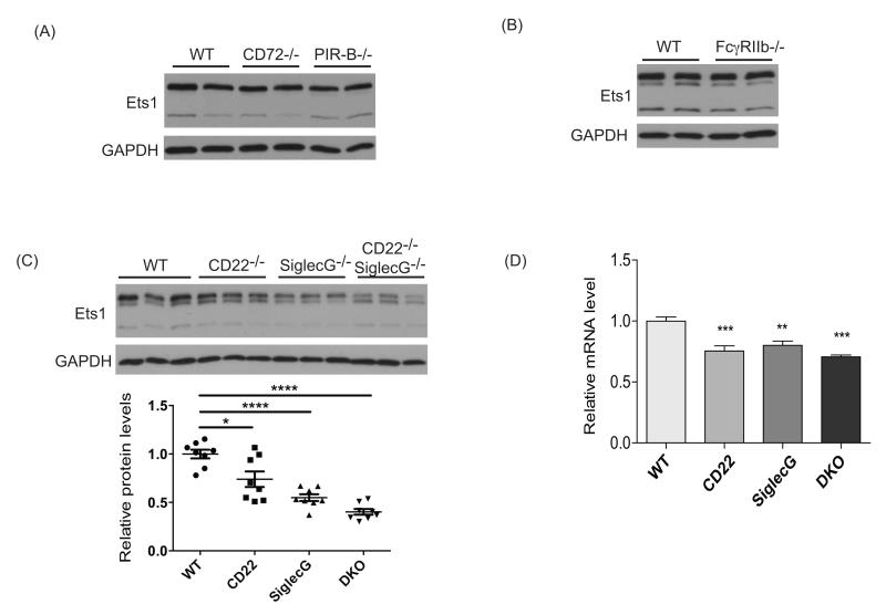
Figure 7. CD22 and SiglecG both contribute to maintaining Ets1 expression in B cells.
(A) Western blot for Ets1 and GAPDH with B cell lysates from wild-type (WT), CD72−/− or PIR-B−/−mice. Shown are results of one of two independent experiments with a total of 4 mice of each genotype being analyzed. (B) Western blot for Ets1 and GAPDH with B cell lysates from wild-type (WT) or FcγRIIb−/− mice. Shown are results of one of two independent experiments with a total of 3 mice of each genotype. (C) Western blot for Ets1 and GAPDH with B cell lysates from wild-type (WT), CD22−/−, SiglecG−/− or CD22−/−SiglecG−/− mice. Shown are the results of one of two independent experiments (n = 8 mice of each genotype). Below is a quantification of multiple Western blots. **p< 0.01, ***p<0.001 (D) RT-qPCR analysis of Ets1 mRNA in B cells from wild-type (WT), CD22−/−, SiglecG−/− or CD22−/−SiglecG−/− B cells (n= 5 for each genotype) ***p< 0.001, ****p<0.0001.

