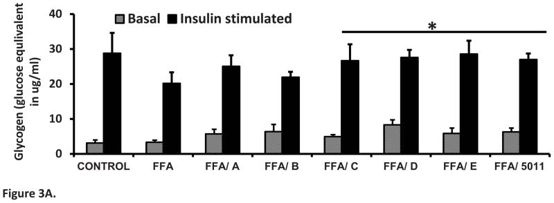
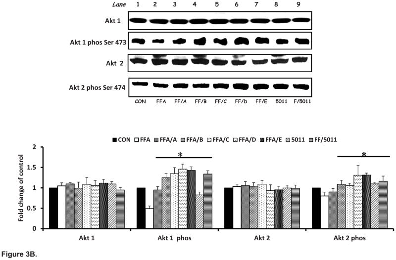
Figure 3. PMI 5011 and isolated compounds enhance glycogen synthesis (A) and Akt Phosphorylation (B) in presence of excess FFAs.

Figure 3A: Rat skeletal muscle cells were treated with 250uM FFAs with or without 10 μg/ml of PMI 5011 or each of the five isolated compounds for 16 hours. Glycogen content was measured by the method of Gomez et al. [12]. PMI 5011 and bioactives C, D and E enhanced insulin stimulated glycogen synthesis to levels not different from that of the control treatment but significantly different from that of the FFA only treatment. Bioactives studied were: davidigenin (A), sakuranetin (B), DMC-1 (C), DMC-2 (D) and synthetic DMC-2 (E). * P < 0.05 vs. FFA only treatment. All experiments were conducted in triplicate.
Figure 3B: Rat skeletal muscle cells were treated with 250uM palmitic acid with or without 10 μg/ml of whole PMI 5011 or each of the isolated compounds for 16 hours before insulin stimulation for 10 minutes. Western blotting showed that palmitic acid attenuated the phosphorylation of Akt-1 and -2 post insulin stimulation, whereas incubation with PMI 5011 or isolated compounds increased Akt phosphorylation significantly when compared to incubation FFA alone (see lane 2 vs. lanes 3 to 9). A) davidigenin, (B) sakuranetin. (C) DMC-1, (D) DMC-2 and E synthetic DMC-2. The bar graphs summarize the results of quantification of gel plots from three experiments. *P < 0.05 vs. FFA only treatment. All experiments were conducted in triplicate.

