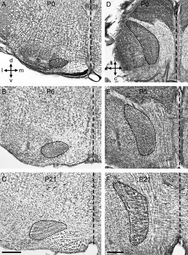
Figure 1.

Anatomical changes in the rat MNTB during postnatal development. A–C: Nissl-stained coronal sections of the rat brainstem at different postnatal ages. D–F: Nissl-stained horizontal sections of the rat brainstem at different postnatal ages. The short-dashed outline represents the MNTB. The long-dashed line represents the midline. MNTB, medial nucleus of the trapezoid body; d, dorsal; v, ventral; l, lateral; m, medial; r, rostral; c, caudal. Scale bars = 500 μm in C (applies to A,B); in F (applies to D,E).
