Figure 3.

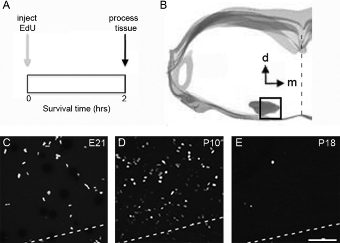
Evidence of cell proliferation in the rat MNTB during postnatal development. A: Experiment design for acute EdU labeling experiments. Rat pups were sacrificed and their brains processed for histological analysis 2 hours after EdU injection (see Materials and Methods). B: Digital tracing outlines the brainstem area used to analyze the density of EdU-labeled cells in coronal sections containing the MNTB. C–E: Exemplar micrographs of EdU-labeled cells at different ages. The dashed line indicates the ventral border of the MNTB. EdU, 5-ethynyl-2'-deoxyuridine; d, dorsal; m, medial; E21, embryonic day 21; P, postnatal day. Scale bar = 75 μm in E (applies to C,D).
