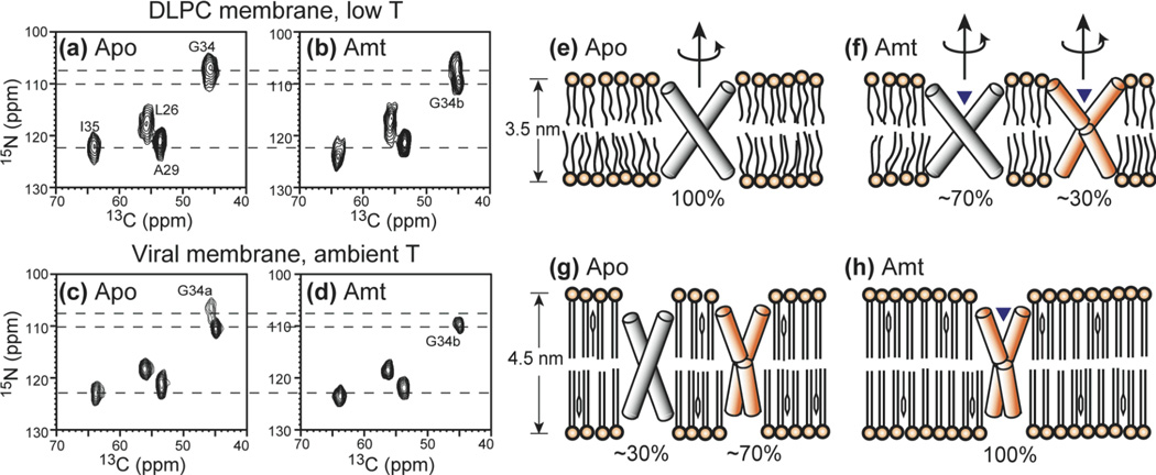
Fig. 6.
2D 15N-13C HETCOR spectra of LAGI AM2-TM in different membranes and drug binding states. (a) Apo AM2-TM in DLPC bilayers at 243 K. (b) Amantadine-bound AM2-TM in DLPC bilayers at 243 K. (c) Apo AM2-TM in viral membranes at 294 K. (d) Amantadine-bound AM2-TM in viral membranes at 303 K. The corresponding helix orientations are shown on the right. (e) Apo peptide in DLPC bilayers. The helices are straight and tilted by 35° from the bilayer normal (37). (f) Amantadine-bound peptide in DLPC bilayers. ~30% of the helices exhibit a kink of 10° at G34. (g) Apo peptide in the viral membrane. ~70% of the helices have the kinked conformation. (h) Amantadine-bound peptide in the viral membrane. All helices have the kinked conformation.

