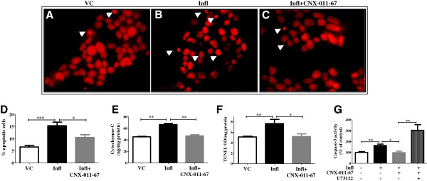
Figure 4.

Chronic GPR40 activation reverses inflammation mediated increase in apoptosis. Apoptotic cells were visualized as condensed nuclei by propidium iodide fluorescence after chronic treatment of NIT1 cells with inflammatory cytokines in presence or ansence of GPR40 agonist (A-D). Representative pictures showing condensed nuclei with white arrow (A-C) demonstrating an increased number of apoptotic cells under inflammation and reversal in apoptotic cells number by GPR40 activation. Thirty images having more than 800 total numbers of nuclei were counted and quantification data were represented by bar graph (D). Inflammation conditions increased and GPR40 agonist decreased cytochrome-c level in rat islets as measured using ELISA assay (E). In NIT1 cells, chronic inflammation increased and GPR40 agonist decreased TUNEL positivity (F). Similarly, inflammation increased caspase-3 activity (G) in NIT1 cells which was reduced by GPR40 agonist. Caspase-3 activity was measured by its ability to cleave DEVD substrate and release of fluorescence. GPR40 induced inhibition of caspae-3 activity was abolished by incubating cells with PLC inhibitor (U73122). (n = 4, *P < 0.05, **P < 0.01, ***P < 0.001).
