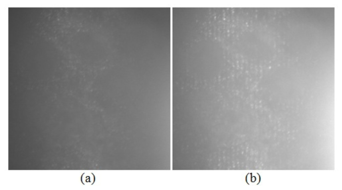
Fig. 2.

Images of phantoms containing SiHa cervical cancer cells labeled with anti-EGFR gold conjugates. The field of view is 54 x 54 μm2. The approximate depth of the imaged optical section is 15-20 μm below the phantom surface. Part (a) shows an inverted widefield reflectance microscope image, and part (b) shows a structured illumination raw image [10].
