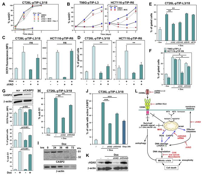
Figure 5. During DICE, DNA DSBs Cause Activation of Caspase-2, Resulting in MOMP.
(A, B) Cell death quantified in the indicated cells after treatment with Dox for up to 7 days in the presence of the indicated inhibitors (zVAD and Nec-1 as in Figure 5C; MVE: 1 μM in CT26L, 0.25 μM in T98G, and 0.2 μM in HCT116 cells). The day 7 data points for CT26L cells are not shown because cells started to die due to overconfluency. P-values were calculated by ANOVA.
(C, D) Quantification of ROS produced (C) and changes in MOMP (D) in cells after knockdown of either CD95L or CD95. Analyses were performed 2 days (CT26L) and 4 days (HCT116) after addition of Dox. P-values were calculated by ANOVA.
(E) Quantification of MOMP in CT26L cells 40 hours after knockdown of CD95L. Cells were pretreated for 1 hour with different caspase inhibitors as indicated. P-values were calculated using t-test.
(F) Quantification of MOMP in cells 3 days (T98G) or 4 days (HCT116) after knockdown of CD95L. Cells were pretreated for 1 hour with different caspase inhibitors as indicated.
(G) From the bottom, DNA degradation, MOMP quantification and ROS staining of CT26L-pTIP-L3 cells transfected with either scrambled (scr) siRNA or a siRNA targeting mouse caspase-2 (100 nM) and treated with Dox for 40 hours. The top panel shows a Western blot for caspase-2 and actin (loading control).
(H) Quantification of DNA degradation in CT26L-pTIP-L3/18 cells 40 hours after knockdown of CD95L. Cells were pretreated for 1 hour with different caspase inhibitors as indicated.
(I) Western blot analysis of caspase-2 and actin in CT26L-pTIP-L3 cells after addition of Dox.
(J) Caspase-2 activity assay of CT26L-pTIP-L3/18 cells 48 hours after knockdown of CD95L. Cells were pretreated with caspase inhibitors where indicated. Etoposide treatment for 24 hours was used as a positive control. P-value was calculated using t-test.
(K) Western blot analysis of γH2AX and actin in CT26L-pTIP-L3/18 cells 40 hours after knockdown of CD95L. Some cells were pretreated with the indicated caspase inhibitors.
(L) Schematic illustrating the DICE subpathway in CT26L cells that results in activation of caspase-2 (blue arrows), which causes MOMP. Cancer cells are protected by the expression of both CD95 and CD95L. For details, see text.
* = p<0.01; ** = p<0.001; *** = p<0.0001; ns = not significant.

