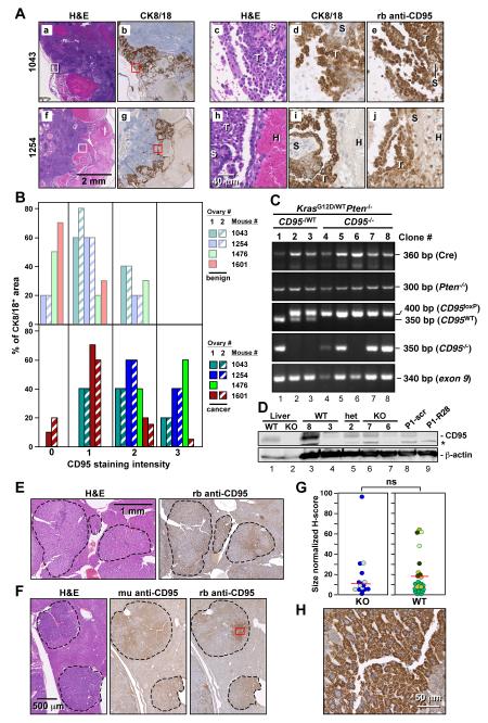
Figure 7. Cancerous Cells Developing from LSL-KrasG12D/wtPtenloxP/loxPCD95loxP/loxPAmhr2-Cre and DEN Treated CD95loxP/loxPAlb-Cre Mice Express CD95.
(A) Immunohistochemistry analysis of sections of ovaries from two LSL-KrasG12D/wtPtenloxP/loxPCD95loxP/loxP mice. a, b, f, and g: larger areas of two ovaries. c, d, e: higher magnifications of H&E, CK8/18 and CD95 staining of the areas boxed in a and b. h, i, j: higher magnifications of H&E, CK8/18 and CD95 staining of the areas boxed in f and g. T, tumor; H, hemorrhage; S, stroma.
(B) Quantification of CD95 staining intensity in ovaries of 4 KrasG12D/wtPten−/− CD95 mutant mice in benign (top) and CK8/18 positive cancerous areas (bottom). Ovary #2 in mouse #1476 had so much infiltration that neither benign nor tumor tissue could be detected. Ovary #2 in mouse #1601 did not contain any benign tissue.
(C) PCR analysis of cloned cell lines derived from mice with ovarian cancer heterozygous or homozygous for CD95loxP. Primers used in the third panel detect the right loxP site in the CD95 gene.
(D) Western blot analysis of single cell clones derived from mice with a genotype of either wild-type, heterozygous, or homozygous deletion of CD95. As a positive control, extracts of a cell line (P1) derived from KrasG12D/wtPten−/− mice (Mullany et al., 2011) are shown infected with a lentiviral shRNA targeting mouse CD95 (R28) or a nontargeting shRNA (scr) as well as liver extracts from wild-type (wt) and CD95loxP/loxPAlb-Cre (ko) mice. *= unspecific band.
(E) H&E and CD95 staining of a liver section of a wild-type mouse 8 months after injection of DEN.
(F) H&E and CD95 staining of a liver section of a CD95loxP/loxPAlb-Cre mouse 8 months after injection of DEN using two different antibodies (a mouse monoclonal (mu) or a rabbit polyclonal (rb) anti-CD95 antibody).
(G) Quantification of CD95 expression in all nodules in sections from two CD95loxP/loxPAlb-Cre and 4 CD95loxP/loxP mice. A total of 15 tumor nodules were analyzed from CD95 knockout livers and 20 nodules were analyzed from wild-type livers. Liver nodules found in individual mice are shown in different colors. ns = not significant.
(H) Magnification of the CD95 stained tumor region in F boxed in red.

