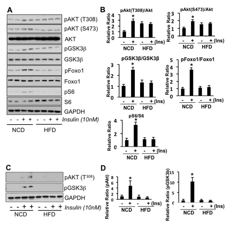
Figure 1.
Representative Western blots of frontal brain tissue from HFD or NCD-fed mice with (+) or without (-) ex vivo insulin stimulation. Mice (n=5-10) were fed with either a 60% HFD for 17 days or 45% HFD for 8 weeks. A) Representative blots from pooled brain tissues of 60% HFD or NCD mice show that insulin stimulation in HFD mouse brain tissue fails to activate IRS-1, Akt, GSK3β, S6 (downstream of mTOR) and Foxo1 (a direct downstream target of Akt). GAPDH served as a loading control. B) Western blot quantitation in 60% HFD vs NCD mice. C) Representative blots from tissue samples from 45% HFD or NCD mice shows HFD mouse brain failure to activate AKT and GSK3 with insulin stimulation. D) Western blot quantitation in 45% HFD vs NCD mice. Student’s t-tests were performed (* p < 0.05).

