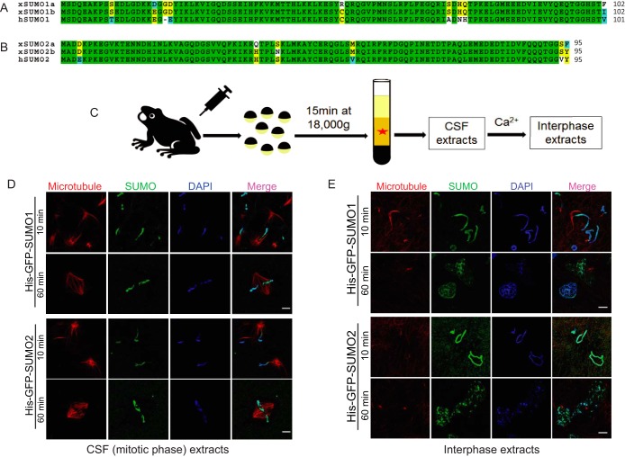
Fig. 1.
SUMO proteins associate with Xenopus sperm chromatin during both mitotic phase and interphase. A, Sequence comparison of Xenopus laevis SUMO1a (NP_001083717), SUMO1b (NP_001090274) and human SUMO1 (NP_003343). Completely conserved residues are shaded green, identical residues are shaded yellow, similar residues are shaded cyan, and different residues are shaded white. B, Sequence comparison of Xenopus laevis SUMO2a (NP_001080085), SUMO2b (NP_001085595) and human SUMO2 (NP_008868). Completely conserved residues are shaded green, identical residues are shaded yellow, similar residues are shaded cyan, and different residues are shaded white. C, CSF-arrested Xenopus egg extract preparation. CSF extracts can be induced to enter interphase by the addition of calcium. D, SUMO proteins associate with sperm chromatin during mitotic phase. Sperm chromatin isolated from male Xenopus was incubated with mitotic Xenopus egg extracts and purified His-GFP-tagged SUMO1 or SUMO2 protein. A strong GFP signal appeared on the chromatin in less than 10 min and persisted for more than 60 min. Microtubule structures formed by rhodamine-labeled tubulin indicate the cell cycle stage of Xenopus egg extracts. DAPI was used for DNA staining. Scale bar equals 10 μm. E, SUMO proteins also associate with sperm chromatin during interphase. The interphase Xenopus egg extract was induced from mitotic egg extract by adding calcium. Sperm chromatin isolated from male Xenopus was incubated with interphase Xenopus egg extract and purified His-GFP-tagged SUMO1 or SUMO2 protein. A strong GFP signal appeared on the chromatin in less than 10 min and persisted even after nuclear formation. Microtubule structures formed by rhodamine-labeled tubulin indicate the cell cycle stage of Xenopus egg extract. DAPI was used for DNA staining. Scale bar equals 10 μm.

