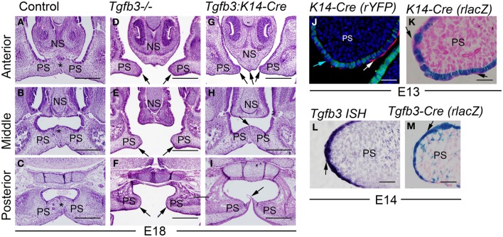
Figure 1.
Milder palatal phenotype of epithelium-specific Tgfb3:K14-Cre mutants than that of Tgfb3 null mutants results from an inability of K14-Cre to recombine in peridermal cells. (A–C) control; (D–F) Tgfb3-/- mutant; (G–I), Tgfb3:K14-Cre (A–I, frontal orientation; all at E18). (A,D,G) on the level of the nasal septum (anterior); (B,E,H), mid-eye level (middle); (C,F,I) on the level of soft palate (posterior). Asterisks in (A–C) indicate confluent midline mesenchyme, black arrows in (D,E) point to unfused palatal shelves, black arrows in (G–I) point to unfused elements of the anterior palate (G), a persistent epithelial seam in the mid-palate (H), and an epithelial bridge in the posterior soft palate (I). (J) a frontal palatal section of a K14-Cre:R26R-YFP embryo at E13; double immuno-fluorescence staining to detect YFP-positive recombined cells (green) and an SSEA1-positive subset of non-recombined peridermal cells (red, white arrow). Light blue arrow points to the DAPI-positive nucleus of a peridermal cell that is SSEA-1-negative and has not been recombined by K14-Cre. (K) A frontal palatal section of a X-Gal-stained K14-Cre:R26R-lacZ embryo at E13, counterstaining with eosin. Black arrows point to apical peridermal cells that were not recombined with K14-Cre. (L) In situ hybridization for Tgfb3 at E14 (palatal frontal section). Black arrow points to a positively staining flattened cell with peridermal appearance. (M) A frontal palatal section of X-Gal-stained Tgfb3-Cre:R26R-lacZ embryo, counterstained with eosin. Black arrow points to an X-Gal-positive flattened cell with peridermal appearance. PS, palatal shelf; NS, nasal septum. Scale bars in (A–I) 200 μm; (J–M) 50 μm.

