Figure 4.
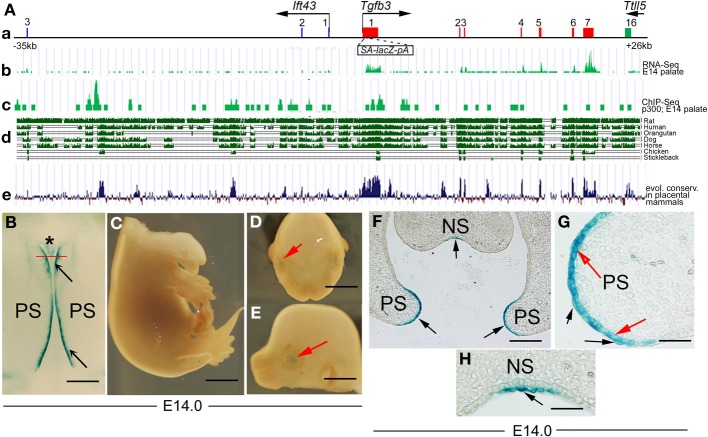
A 61-kb genomic region including theTgfb3 gene targets reporter activity to the MEE and adjacent periderm. (A) Schematic representation of the 61-kb region (see main text) includes (a) the Tgfb3 gene (exons in red); exons 1–3 of the Ift43 gene (blue boxes) and exon 16 of the Ttll5 gene (green box). That Tgfb3 but neither of its neighbors is actively expressed at E14.5 is shown in (b), RNA-seq profile in mouse palate at E14.5—(FaceBase Enhancer Project; A Visel). Different types of sequence analysis are suggestive of possible enhancer sites: (c) p300 Chip-seq profile in mouse palate at E14.5 (ref: FaceBase Enhancer Project; A Visel), (d) evolutionary sequence conservation among selected vertebrate species in this sequence (UCSC genome browser) and (e) evolutionary sequence conservation among placental mammals (UCSC genome browser). (B–E) 61-kb BAC-lacZ embryos showing β-gal activity (blue staining) in tips of palatal shelves (B, black arrows), nasal septum (B, asterisk), and lens (D,E, red arrows). (B) Roof of mouth at E14, inferior view, anterior on the top; (C) torso at E14, right lateral view; (D) head (mandible removed), left lateral view; (E) head (mandible removed), frontal view. (F–H) Frontal sections of an X-Gal stained 61-kb lacZ-BAC embryo at the level of nasal septum (indicated by the red line in B). (F) X-Gal staining (black arrows) at the tips of the pre-fusion palatal shelves (PS) and in the nasal septum (NS) is present in cells of both the basal MEE layer (red arrows in G) and the overlying periderm layer (black arrows in G), and periderm in the nasal septum (black arrow in H). Scale bars in (B), 500 μm; (C–E), 1 mm; (F) 200 μm; (G,H), 50 μm.

