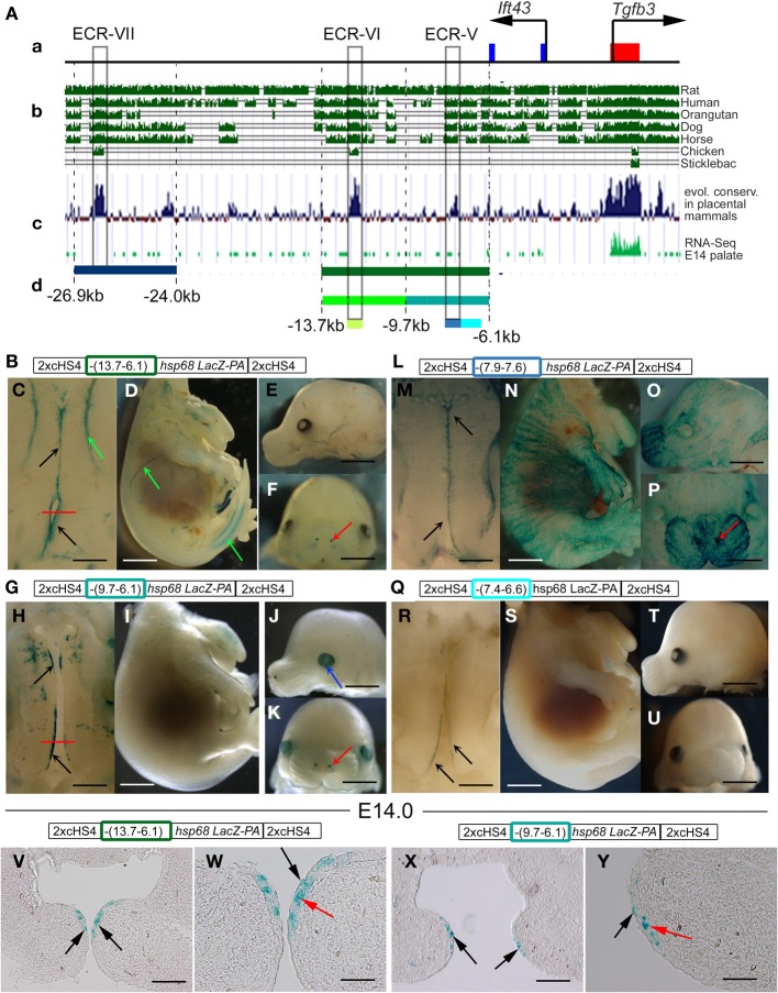
Figure 6.
Cis-regulatory elements targeting reporter activity to the MEE and adjacent periderm are located in intron 2 of the upstream Ift43 gene. (A) Schematic representation of a 28-kb sub-region of the 61-kb genomic fragment (a, see Figure 4 and the main text) that includes Tgfb3 exon 1 (red box) and exons 1 and 2 of the Ift43 gene (blue boxes), aligned with (b) evolutionary sequence conservation among selected vertebrate species (UCSC genome browser), and (c) among placental mammals (UCSC genome browser), and (d) RNA-seq profile in mouse palate at E14.5 (FaceBase Enhancer Project; A Visel) used to identify the positions of non-coding evolutionary conserved regions (ECRs-V, -VI, and -VII: gray boxes). Colored bars below (d) correspond to DNA fragments (see also Figure 3) examined by transient transgenic reporter assay in constructs shown schematically above images of the stained embryos generated (B–Y). (C–F) Transgenic reporter embryos carrying a 7.6-kb DNA fragment from −13.7 to −6.1 kb (green bar below (c) and green rectangle in construct schematic) showing β-gal activity (blue staining) in tips of palatal shelves (C, black arrows), blood vessels (C,D, green arrows) and nostrils (F, red arrow). (H–K) Transgenic reporter embryos carrying a 3.6-kb DNA fragment from −9.7 to −6.1 kb (blue-green bar below (c) and blue-green rectangle in construct schematic) showing β-gal activity (blue staining) in tips of palatal shelves (H, black arrows), lens (J, blue arrow) and nostrils (K, red arrow). (M–P) Transgenic reporter embryos carrying a 0.3-kb DNA fragment (ECR-V) from −7.9 to −7.6 kb [light blue bar below (c) and light blue rectangle in construct schematic] showing β-gal activity (blue staining) in tips of palatal shelves (M, black arrows), apical ectoderm (N–P) and nostrils (P, red arrow). (R–U) Transgenic reporter embryos carrying a 0.8-kb DNA fragment from −7.4 to −6.6 kb (turquoise bar below (c) and rectangle in construct schematic) showing β-gal activity (blue staining) in tips of posterior palatal shelves (R, black arrows). (V–W) Frontal sections of the X-Gal-stained 7.6-kb fragment transgenic embryo shown in (C) at the level of the posterior palate (indicated by the red line in C). Staining at the tips of palatal shelves (black arrows in V) is in both MEE cells (red arrow in W) and periderm cells (black arrow in W). (X,Y) Frontal sections of the X-Gal-stained 3.6-kb fragment transgenic embryo shown in (H) at the level of the posterior palate (indicated by the red line in H). Staining at the tips of palatal shelves (black arrows in X) is in both MEE cells (red arrow in Y) and periderm cells (black arrow in Y). Scale bars in C,H,M,R, 500 μm; D,I,N,S,E,F,J,K,O,P,T,U, 1 mm; V,X, 100 μm; W,Y, 50 μm.

