Figure 1.

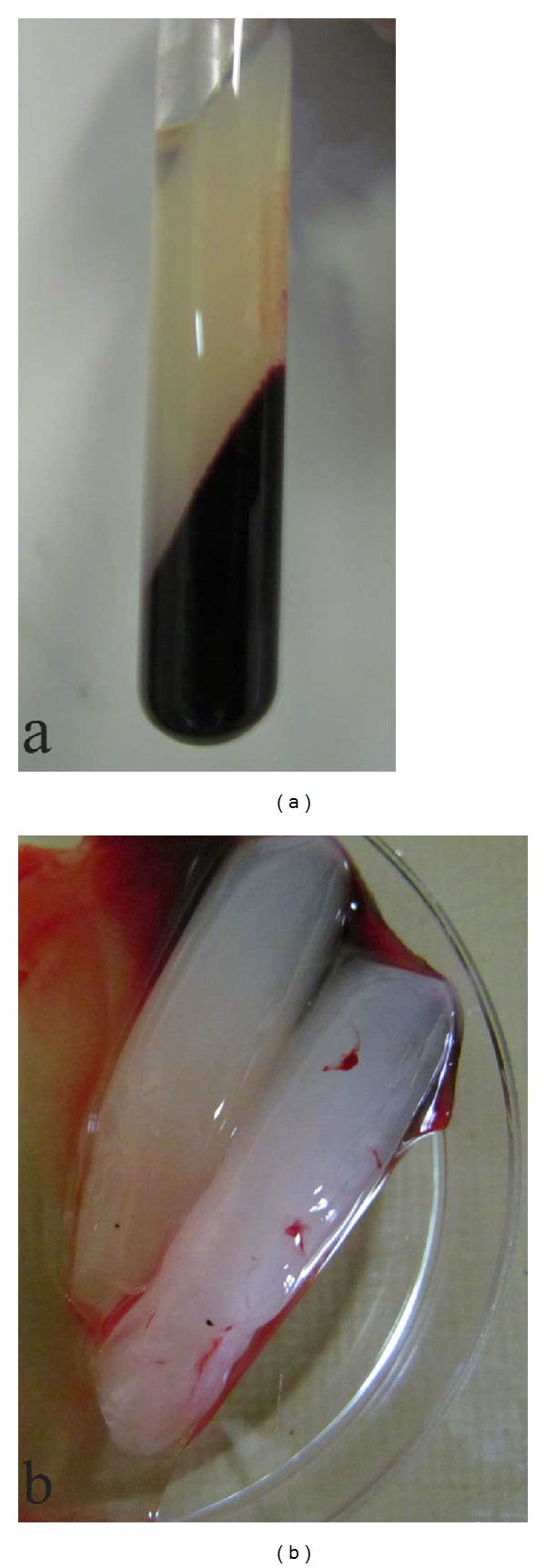
Venous blood sample following centrifugation indicating the PRF clot in the middle layer of the test tube (a) and the removed clots placed inside sterile petri dish (b).

Venous blood sample following centrifugation indicating the PRF clot in the middle layer of the test tube (a) and the removed clots placed inside sterile petri dish (b).