
Figure 7.
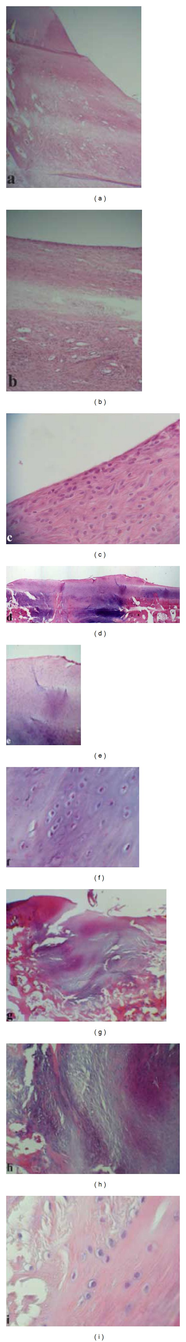
Histologic sections of the representative defects from the PRF treated group at different time intervals: 4 weeks ((a)–(c)), 16 weeks ((d)–(f)), and 24 weeks ((g)–(i)). Haematoxylin-eosin staining; original magnification: 40x ((a), (d), and (g)), 100x ((b), (e), and (h)), and 400x ((c), (f), and (i)).