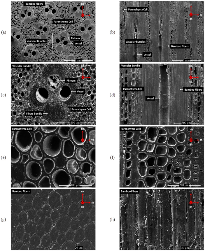
Figure 1. SEM micrographs of the raw bamboo culm with different constituents.
zoom-in views of bamboo's vascular bundles along with the parenchyma ground and bamboo fibers along the transversal ((a), (c), (e) and (g)) and longitudinal ((b), (d), (f) and (h)) directions. As displayed, fibers and parenchyma cells, comparably, possess the majority of bamboo culm whereas vessels possess less contribution.

