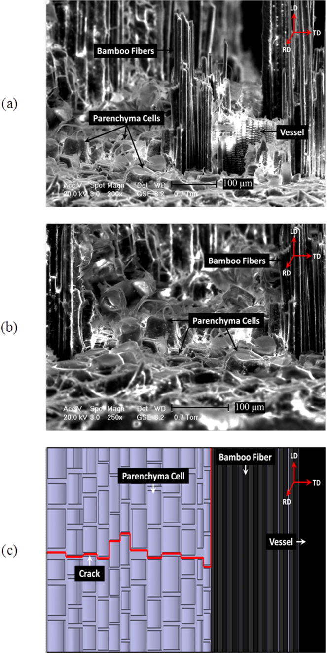
Figure 4.
Crack propagation in bamboo culm's three-dimensional (3D) structure revealed by a tensile fractured sample: (a–b) ESEM micrographs displaying the tensile fracture surfaces of the bamboo culm at high magnification, with focus on the (a) fibers pull-out and the (b) intact parenchyma cells; (c) schematic representation of crack growth within bamboo's different constituents from the view of longitudinal direction (LD).

