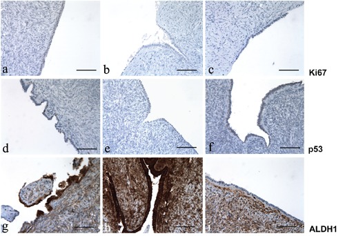
Figure 1.

Ki67, p53 and ALDH1 immunoreactivity in ovary; left column, after treating with tamoxifen; center column, after treating with clomid; right column, untreated control group. a-c) Immunoreactivity for Ki67; note the absence of Ki67 expression in all the three representative groups. d-f) Immunoreactivity for p53; p53 expression is low in the ovarian epithelium exposed to tamoxifen; in contrast, there is no p53 expression in ovary exposed to clomid and in the control group. g-i) immunoreactivity for ALDH1; the expression of ALDH1 is strong in ovaries exposed to tamoxifen and clomid, whereas no immunopositivity for ALDH1 was observed in the control group; intense staining for ALDH1 was noted in stroma (internal control). Scale bars: 100 µm.
