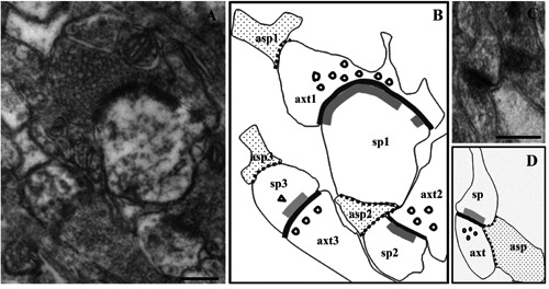
Figure 1.

Morphometric analyses of axo-spinous synapses and glia-synapse relationships. Examples of micrographs from CA1 stratum radiatum used for morphometrical analyses, showing axo-spinous synapses surrounded by distal astrocytic processes (A and C). Schematic drawings reproducing the corresponding micrographs are depicted in B and D. A and B, an axon terminal (axt1) contacted extra-synaptically (i.e. far away from the cleft) by an astrocytic process (asp1), and two dendritic spines (sp1 and sp3) contacted by extra-synaptic glial processes (asp2 and asp3) are shown; asp2, extra-synaptic on sp1, is also in direct apposition with axt2/sp2 bouton-spine interface, thus being intra-synaptic on this synapse. In C and D, a synapse is contacted at its bouton-spine interface by an astrocytic process (intra-synaptic) covering both pre- and post-synaptic profiles. The membrane apposition between neuronal synaptic profiles and glial processes is marked with dotted lines; bouton-spine interfaces (bold black line) and postsynaptic specializations (double bold gray line) are also marked. As an example of morphometric analyses carried out in the study, measures calculated for axt1/sp1 synapse (in B) are as follows: axt1 perimeter, 2396 nm; portion of axt1 profile covered by asp1, 238 nm (glia-covered/total profile ratio: 0.1); sp1 perimeter, 1310 nm; portion of sp1 profile covered by asp2, 332 nm (glia-covered/total profile ratio: 0.25); length of post-synaptic specializations in sp1, 585 nm and 67 nm; length of axt1/sp1 bouton-spine apposition, 800 nm. sp, dendritic spine; axt, axon terminal; asp, astrocytic process. Scale bars: A,C) 250 nm.
