Figure 2.

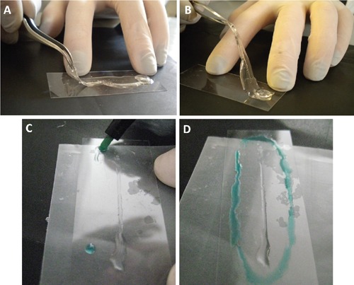
A-B) PDMS peeled from the glass. C-D) The area with cells attached to the glass is outlined with an hydrophobic barrier pen (in green) to allow the immunostaining.

A-B) PDMS peeled from the glass. C-D) The area with cells attached to the glass is outlined with an hydrophobic barrier pen (in green) to allow the immunostaining.