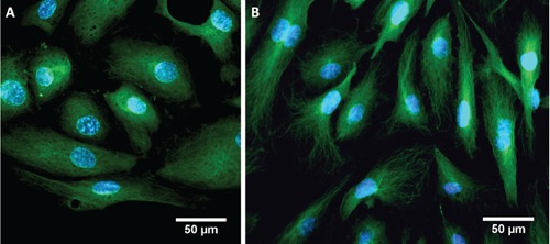
Figure 4.

Immunofluorescence of HUVEC cells: nuclei are stained in blue, using DAPI fluorescent dye; α/β tubulin is in green, in flow conditions in MFD with flow rate of 2 µL min–1 for the first 20 h and then 30 µL min–1 for 2 h (A) and in static conditions in multiwells (B).
