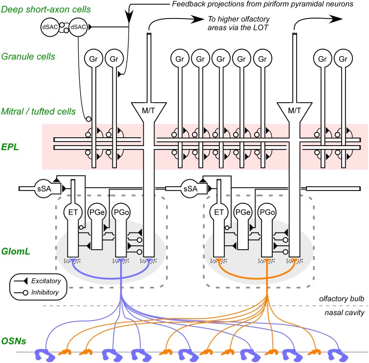
Figure 1.
Circuit diagram of the mammalian olfactory bulb. The axons of olfactory sensory neurons (OSNs) expressing the same odorant receptor type converge and arborize together to form glomeruli (shaded ovals; two depicted) on the surface layer of the olfactory bulb. Intrinsic OB interneurons innervate each glomerulus, including olfactory nerve driven periglomerular cells (PGo), external tufted cell-driven periglomerular cells (PGe), and external tufted cells (ET). Superficial short-axon cells (sSA), closely related to PG cells and possibly part of the same heterogeneous population, are not associated with specific glomeruli but project broadly and laterally within the deep glomerular layer. Principal neurons include mitral cells and tufted cells (collectively depicted as M/T), which interact via reciprocal connections in the external plexiform layer (EPL) with the dendrites of inhibitory granule cells (Gr), thereby receiving recurrent and lateral inhibition, and project out of the OB to several regions of the brain. The heterogeneous deep short-axon cell population (dSAC) includes cells that deliver GABAergic inhibition onto granule cells and one another, and, along with granule cells, receive centrifugal cortical input from piriform pyramidal cells. OE, olfactory epithelium (in the nasal cavity); GL, glomerular layer; EPL, external plexiform layer; MCL, mitral cell layer; IPL, internal plexiform layer; GCL, granule cell layer. Filled triangles denote excitatory synapses; open circles denote inhibitory synapses. Speckles surrounding OSN terminals denote volume-released GABA and dopamine approaching presynaptic GABAB and dopamine D2 receptors. Note that sSA-PG and sSA-sSA synapses are depicted as excitatory despite being GABAergic (discussed in Cleland, 2014). Figure adapted from Cleland et al. (2012).

