
Figure 1.
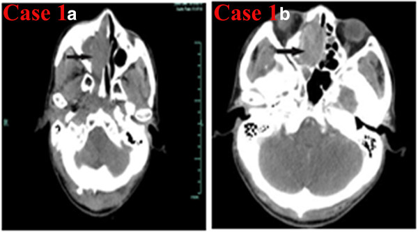
CT of the sinonasal small-cell neuroendocrine carcinoma of Case 1. a) Noncontrast axial CT showing a soft-tissue mass in the right maxillary sinus involving the right nasal cavity, right ethmoid sinus, and sphenoid sinus. b) Contrast-enhanced CT showing that the lesion was strongly enhanced.