
Figure 4.
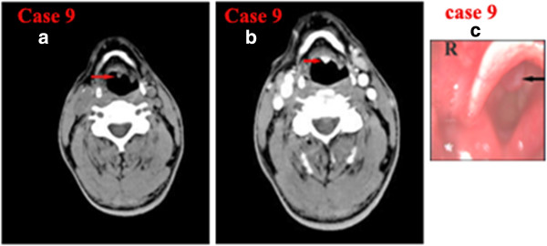
CT of laryngeal small-cell neuroendocrine carcinoma of Case 9. a) Noncontrast axial CT showing a small well-defined soft-tissue mass in the left epiglottic base not involving the bilateral vocal cord and laryngeal cartilages. b) Contrast-enhanced CT showing that the lesion was strongly enhanced. c) Laryngostroscopy showing a red smooth mass in the laryngeal surface of epiglottis.