Figure 4.
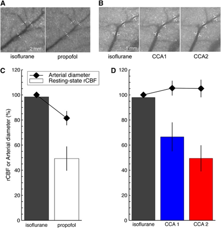
Regional cerebral blood flow (rCBF) and microvascular tone. Close-up on the middle cerebral artery (MCA) before induction of the cortical spreading depolarization (CSD) under different anesthesia regimens, isoflurane and propofol (A), or under different upstream blood supply conditions (B). These images show speckle contrast temporally averaged over 40 seconds. Sequential common carotid artery (CCA) ligations were performed under isoflurane. Common carotid artery 1 (CCA1) refers to the ligation of the left CCA. Common carotid artery 2 (CCA2) refers to the ligation of both CCAs. Three segments along the distal portion of the MCA were used to quantify arterial diameter changes, as shown by the white markers. Resting-state rCBF (bars, n=5 animals) and arterial diameter (diamonds) when the anesthesia regimen was modified (C) or when the upstream blood supply to the brain was altered (D). Resting-state rCBF was the averaged rCBF value before needle prick calculated across n=5 animals from group 1 (C) or across n=5 animals from group 3 (D). Regional cerebral blood flow 100% refers to isoflurane rCBF under normal upstream blood supply. Similarly, the arterial diameter measured under isoflurane at normal blood supply before needle prick was set to 100%. Changes from this 100% arterial diameter are reported as mean±s.d. for the three segments of the MCA (shown in A and B). Note that the region of interest for rCBF calculation was intentionally not placed on a major blood vessel to reflect tissue rCBF. Microvascular tone and resting-state rCBF were closely related under normal blood supply to the brain but dissociated when the CCAs were ligated and upstream blood supply reduced.

