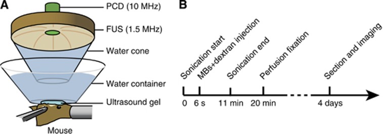
Figure 1.
(A) Schematic illustration of the experimental setup. A 1.5-MHz single element focused ultrasound (FUS) transducer was used for blood–brain barrier opening. It was confocally aligned with a transducer for passive cavitation detection (PCD). The left hippocampus of each mouse was targeted during sonication and the right hippocampus was used as the control. (B) Illustration of the experimental timeline. Sonication started ∼6 seconds before the injection of a mixture of Definity microbubbles and one dextran with a molecular weight of 3, 70, 500 or 2,000 kDa. During the 11-minute sonication, cavitation detection was performed. The mice were transcardially perfused and fixed at ∼20 minutes after dextran injection and then the brain tissue was sectioned for fluorescence imaging.

