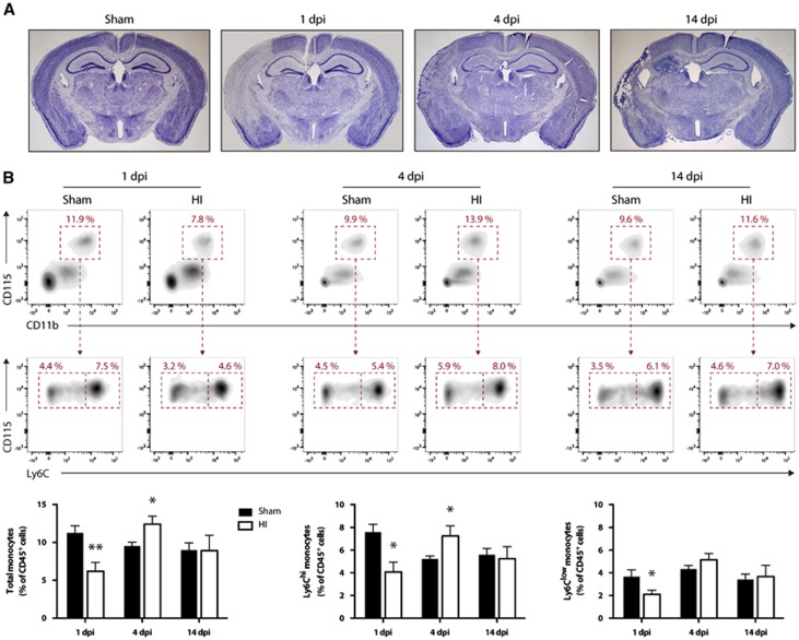
Figure 1.
Hypoxic-ischemic-induced brain infarction and modulation of blood monocytes levels. Hypoxia-ischemia (HI) was induced in 6-month-old male mice by the permanent occlusion of the right common carotid artery followed by exposure to hypoxia (8% O2, 92% N2) for 30 minutes. (A) Representative photomicrographs of Nissl-stained coronal brain sections at different time points after HI. There is marked neuronal loss starting at 1 day post injury (d.p.i.) and a progressive cerebral atrophy of the ipsilateral hemisphere (left) compared with the noninjured contralateral side (right). (B) Flow cytometric analysis of blood monocytes after HI. Monocytes were identified by the expression of the following surface antigens: CD45+CD11b+CD115+Ly6G- whereas Ly6Chi monocytes and Ly6Clow monocytes subsets were separated based on their respective expression level of Ly6C (high versus low). (Results are expressed as means±s.e.m.; n=5 to 7. *P<0.05, **P<0.01 (versus sham); multiple Student's t-test).

