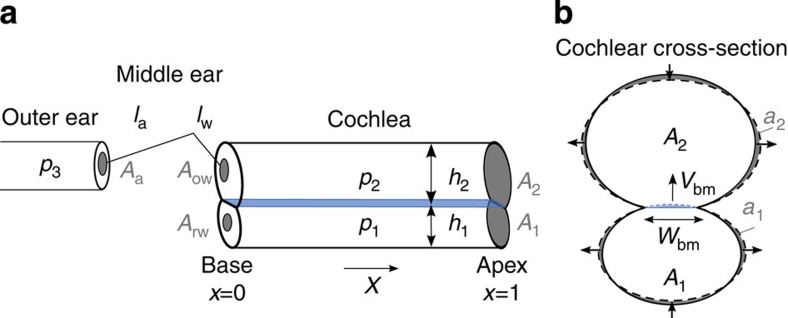
Figure 1. Anatomy of the ear.
(a) Sound causes a pressure vibration p3 in the ear canal and a motion of the ear drum (area Aa). The middle ear’s ossicles, namely the mallus of length la, incus of length lw, and stapes convey the motion to the inner ear, or cochlea, to vibrate the oval window (area Aow) and the round window (area Arw). The pressures p1 in the scala tympani and p2 in the scala vestibuli change accordingly. (b) A transverse section of the inner ear shows the basilar membrane separating two chambers of cross-sectional area A1 and A2. Vibration of the membrane (velocity Vbm) and deformation of the cochlear bone, at constant circumference, lead to area changes a1 and a2.

