
Figure 3.
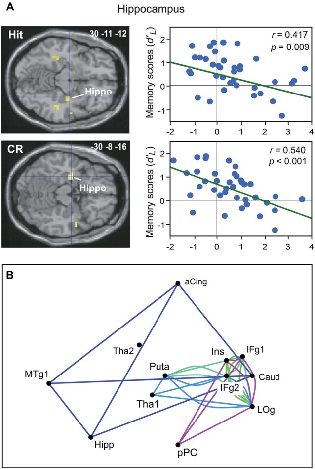
Neural basis of odor recognition memory. (A) Decreased activation intensity in the right and left hippocampus as a function of memory scores (d’L) for Hit and CR in all participants (adapted from Royet et al., 2011). (B) The module in dark blue shows four regions functionally connected during the Hit condition. Other modules were also found during the CR, Miss or False alarm conditions. aCing, anterior cingulate; Caud, caudate nucleus; Hipp, hippocampus; IFg, Inferior frontal gyrus; Ins, insula; LOg, lateral orbital gyrus; MTg, medial temporal gyrus; pPC, posterior piriform cortex; Puta, putamen; Tha, thalamus (adapted from Meunier et al., 2014).