
Fig. 3.
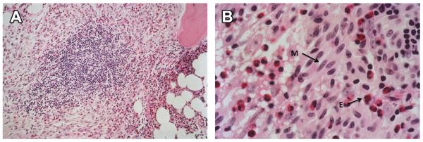
(A) Bone marrow biopsy in systemic mastocytosis, intermediate magnification (Hematoxylin and Eosin, 100×) featuring atypical, spindle-shaped mast cells and intermingled eosinophils surrounding a lymphoid aggregate. (B) Bone marrow biopsy in systemic mastocytosis, high magnification (Hematoxylin and Eosin, 400×) showing atypical, spindle-shaped mast cells (M) with numerous intermingled eosinophils (E). (Courtesy of Charles W. Ross, MD, University of Michigan, Ann Arbor, MI.)