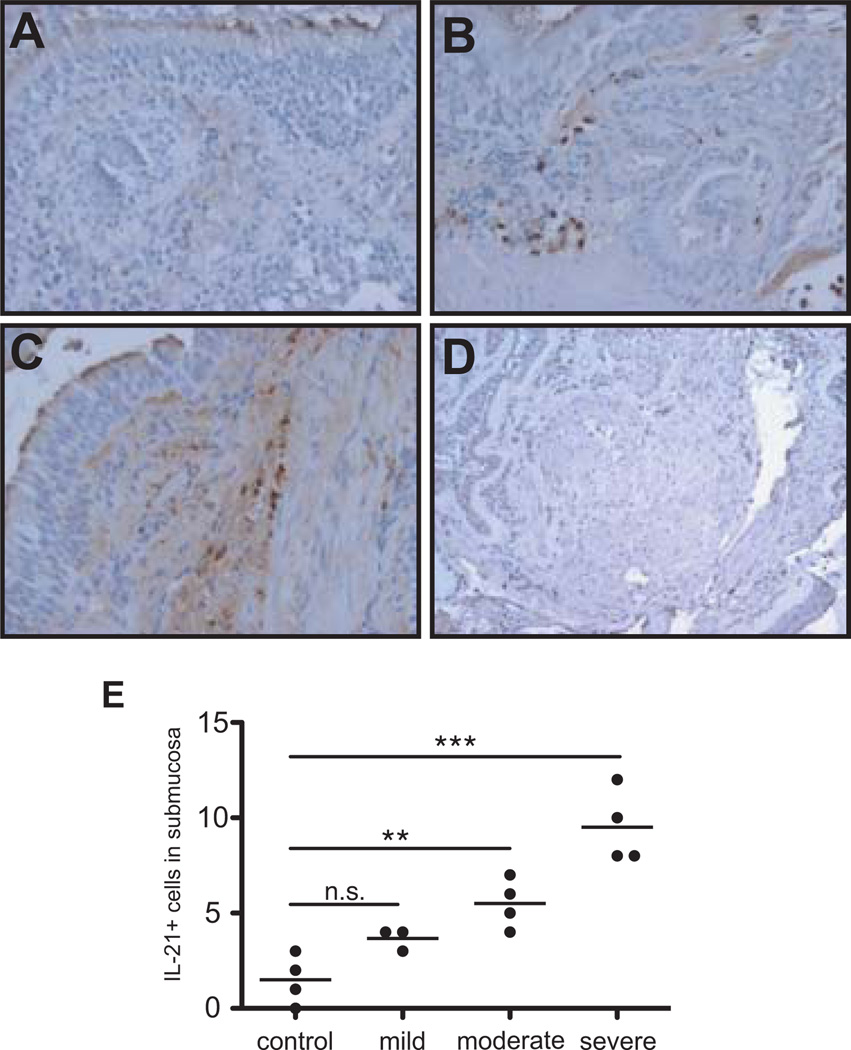
Figure 3. IL-21 expression is increased in human asthmatic airways.
Immunohistochemical analysis for IL-21 was conducted on sections from bronchial biopsies obtained from non-asthmatic (A), moderate asthmatic (B), and severe asthmatic (C) individuals (20× magnification). A section from a severe patient was also stained with an isotype control (D). Quantification of the IL-21+ cells/field in the subepithelium is presented in panel E (*p < 0.05, **p < 0.01).

