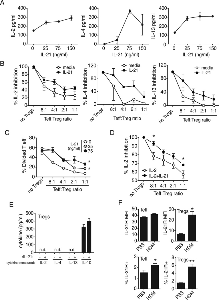
Figure 4. IL-21 overrides Treg-mediated suppression of T effector cytokine production and proliferation.
CFSE-labeled effector T cells (CD4+CD25−) derived from lymph nodes were stimulated with anti-CD3 and anti-CD28 and treated with IL-21 in the presence of increasing numbers of Tregs (CD4+CD25bright). After 5 days, (A, B) supernatants were collected for cytokine analysis, and (C) cells harvested for detection of proliferation by CFSE dilution. Proliferation is expressed as % divided T cells and expressed as the mean ± SEM of three wells. Effector T cells (CD4+CD25−) derived from lymph nodes were in were incubated with increasing numbers of IL-2 or IL-2+IL-21-pretreated Tregs (CD4+CD25bright), after 5 days supernatants were collected for IL-2 analysis (D). Supernatants from CD4+CD25bright cells (5.0×10⁴/well) were collected for cytokine analysis, from media or IL-21-treated Tregs, data is expressed as the mean ± SEM of three wells (C). The influx of IL-21R+ Tregs (CD4+CD25+Foxp3+) in the lungs is greater than IL-21R+ effector T cells after HDM challenge. Tregs also strongly upregulate the IL-21R on their surface (MFI) compared to CD4+ effector T cells following allergen exposure (F), data is expressed as the mean ± SEM of 4–8 mice (*p < 0.05, **p < 0.01). Data is representative of 2–5 independent experiments.

