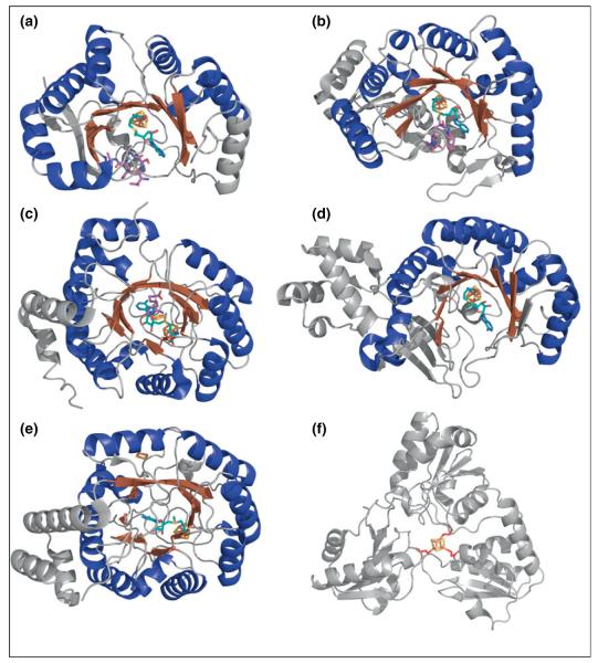
Figure 2.
Crystal structures of representative RS enzymes and Dph2. The Fe–S clusters are shown in rust for iron and yellow for sulfur. SAM molecules (teal carbons) and substrates (purple carbons) are also shown. (a) PFL-AE with SAM and the substrate PFL peptide (PDB ID: 3CB8). (b) SPL with SAM and substrate dinucleoside 5R-SP (PDB ID: 4FHD). (c) BioB with SAM, DTB substrate, and the additional [2Fe–2S] cluster (PDB ID: 1R30). (d) RlmN with SAM (PDB ID: 3RFA). (e) HydE with SAM and additional [2Fe–2S] cluster (PDB ID: 3IIZ). (f) Dph2 with the iron coordinating cysteines in red (PDB ID: 3LZD).

