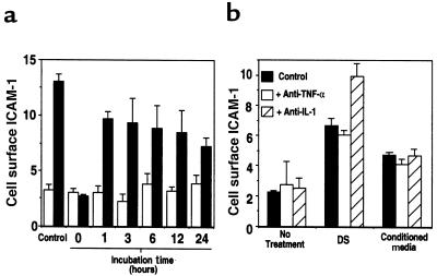
Figure 7.
DS initiates autocrine induction of cell surface ICAM-1. Cell surface ICAM-1 was measured on human dermal microvascular endothelial cells by ELISA, as described in Methods. (a) Cells were cultured for the indicated times in media (open bars) or media containing 50 μg/mL DS (filled bars). These media were then depleted of DS by anion exchange, followed by addition to fresh cells for 24 hours. Bars labeled “control” represent cells cultured continuously for 24 hours. (b) Effect of neutralizing antibodies to TNF-α or IL-1 on ICAM-1 induction by media alone (no treatment), 50 μg/mL DS, or conditioned media from cells treated for 24 hours with DS and then depleted of DS as described in a (conditioned media). Media were pretreated in the absence or presence of 5 μg/mL anti–TNF-α or 10 μg/mL anti–IL-1 and then added to endothelial cell culture for 24 hours before assay. Data represent mean relative cell surface ICAM-1 expressed as mean OD/min ± SD for triplicate determinations.

