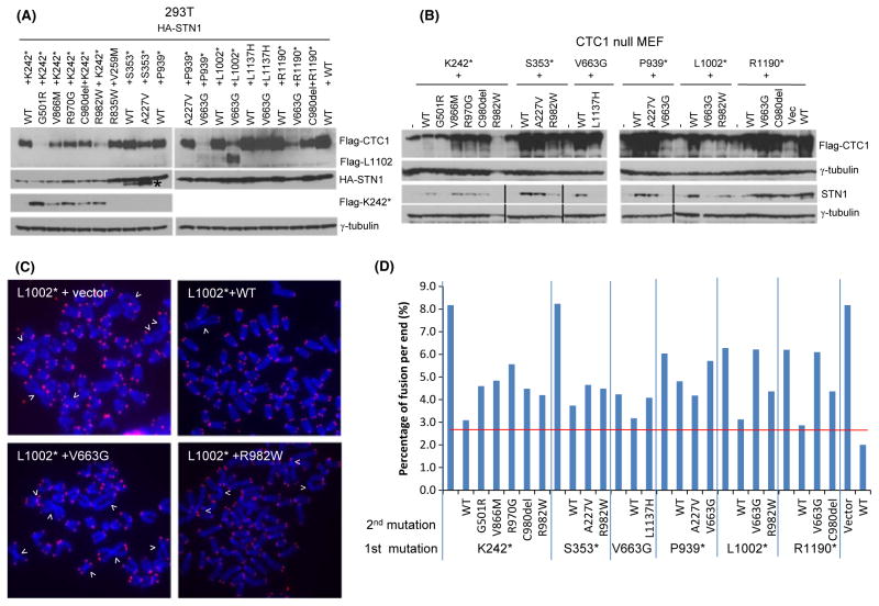
Fig. 4.
Characterization of CTC1 compound heterozygous mutations in vivo. (A) Lysates from 293T cells stably expressing HA-STN1 and the indicated Flag-CTC1 mutations were probed with anti-Flag and anti-HA antibodies. γ-tubulin served as loading control. (B) Flag-tagged WT and mutant CTC1 were expressed in CTC1−/− MEFs and lysates probed with antibodies against Flag and STN1. Dual expression of mutant CTC1 in CTC1−/− MEFs was performed as described in Experimental procedures. γ-tubulin served as loading control. (C) Telomere PNA-FISH and DAPI analysis on metaphase chromosome spreads of CTC1−/− MEFs stably expressing either WT or compound heterozygous CTC1 mutants indicated in (B), using Tam-OO-(CCCTAA)4 telomere peptide nucleic acid (red) and DAPI (blue). White arrowheads: fused chromosomes. (D) Quantification of chromosome fusions in (C). A minimum of 30 metaphases were examined. Red horizontal line refers to average number of chromosome fusions from seven independent experiments of CTC1 mutant cell lines co-expressing WT CTC1.

