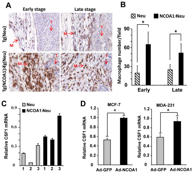
Fig. 2. NCOA1 overexpression in mammary tumor cells enhances macrophage recruitment and CSF1 expression.
A. Immunostaining of macrophages (brown color) with F4/80 antibody in Tg(Neu) (Neu) and Tg(NCOA1)×Tg(Neu) (NCOA1×Neu) mammary tumors. T, tumor area; M, macrophage. B. Macrophage numbers in Tg(Neu) and Tg(NCOA1)×Tg(Neu) tumors at early and late stages. Macrophages in 10-viewing fields at 400× magnification were counted on each tumor section. Five tumors were analyzed for each group. Data are presented as mean ± standard deviation (SD). *, p<0.05. C. qPCR measurement of CSF1 mRNA levels in individual Tg(Neu) and Tg(NCOA1)×Tg(Neu) mammary tumors. D. Adenovirus-mediated NCOA1 expression (Ad-NCOA1) in MCF-7 and MDA-MB-231 cells induced CSF1 expression as assayed by qPCR. Adenovirus-mediated GFP expression (Ad-GFP) served as a control.

