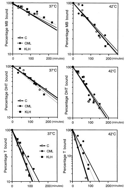
Figure 2.
Dissociation kinetics of ARs in genital skin fibroblasts. Normal (C, open circles and bold lines) and mutant (CML, filled circles and normal lines; KLH, filled squares and thin lines) fibroblast monolayers were exposed to 2 nM [3H]MB (top), 3 nM [3H]DHT (middle), or 3 nM [3H]T (bottom) at 37°C (left) or 42°C (right) for 2 hours. The radiolabeled medium was discarded and replaced with one containing 200-fold excess unlabeled androgen; replicate samples were removed at the indicated times and assayed for [3H]androgen that was still receptor bound. Each data point, the mean of 4 replicates, is expressed as a percentage of maximum binding at time 0. Vertical axes are on the same logarithmic scale, except for bottom right panel.

