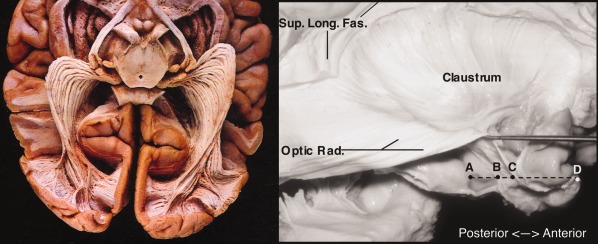
Figure 3.

The optic radiations (ex vivo samples). Left: inferior view. Basal structures, cortex, and white matter removed. From: Gluhbegovic & Williams, The Human Brain (1980). With permission of Lippincott Williams & Wilkins, 1980. Right: lateral view of optic radiation (Optic Rad.) with overlying structures removed. Note continuous, sheet‐like structure rather than three spatially discrete bundles. Also labeled: superior longitudinal fasciculus; A, hippocampal head; B, tip of temporal horn of lateral ventricle; C, anterior tip of optic radiation; D, temporal pole. From Rubino, Rhoton, Tong, De Oliviera (2005) Three‐dimensional Relationships of The Optic Radiation. Neurosurgery 57 (Sup. 4), p227, with permission Wolters Kluwer Health.
