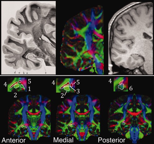
Figure 5.

Illustration of lateral geniculate nucleus (LGN) candidate seed regions with reference to the region's known anatomy. Top row: coronal LGN and radiations in section from Meyer [2012; left] at level of LGN. e, external geniculate body (LGN); a, tapetum; b, internal sagittal marrow; c, external sagittal marrow; d, subcortical marrow. Approximately equivalent image on diffusion‐weighted (center) and T1 (right) images. Bottom row: coronal DWI images with LGN regions of interest illustrated on a mirrored hemisphere at relatively anterior, medial and posterior regions (inset: enlargement of LGN region). Numbers refer to the six regions of interest described in text.
