
Fig. 1.
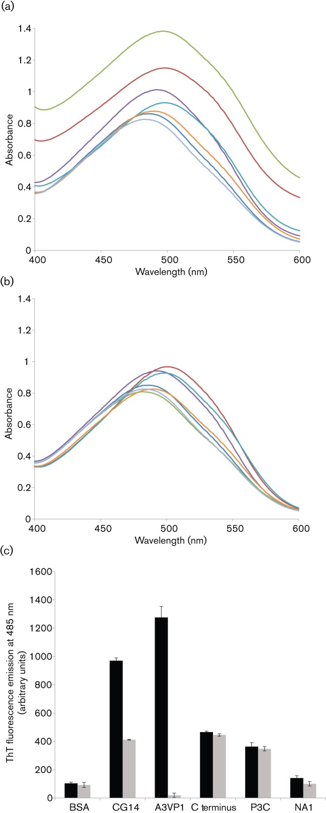
Binding of amyloidophilic dyes to purified recombinant P1 polypeptides. All samples were tested at 5 µM. (a) Absorbance spectra of CR alone and in the presence of aggregated (stirred) samples. (b) Absorbance spectra of CR alone and in the presence of unstirred samples. Dark blue, BSA; red, CG14; green, A3VP1; purple, C terminus; aqua, P3C; orange, NA1; light blue, CR alone. (c) ThT-dependent fluorescence of stirred and unstirred samples. ThT fluorescence emission at 485 nm, following excitation at 442 nm, is shown. P1 alone did not contribute to the fluorescence signal. No change in ThT fluorescence intensity was observed in the absence of P1. Black bars, stirred; grey bars, not stirred. Error bars are sem.