
Fig. 7.
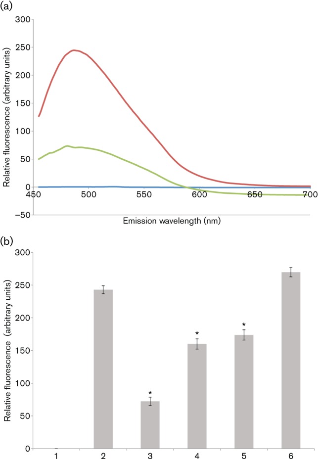
EGCG inhibits amyloid fibrillization by S. mutans extracellular proteins. (a) ThT fluorescence emission spectra, following excitation at 442 nm. S. mutans extracellular proteins were stirred in the cold to induce amyloid fibrillization in the presence (green) and absence (red) of 1 mM EGCG and ThT fluorescence emission was monitored from 450–700 nm. Background fluorescence was evaluated in the absence of added protein (blue). (b) Inhibition of amyloid fibrillization tested over a range of EGCG concentrations. ThT fluorescence emission at 485 nm, following excitation at 442 nm, is shown. Bars: 1, ThT-only control; 2, no EGCG; 3, 1 mM EGCG (P≤0.001); 4, 100 µM EGCG (P≤0.0011); 5, 10 µM EGCG (P≤0.0024); 6, 1 µM EGCG. Error bars represent sem. Significant differences between samples with and without inhibitor, indicated above in parentheses, are denoted with asterisks.