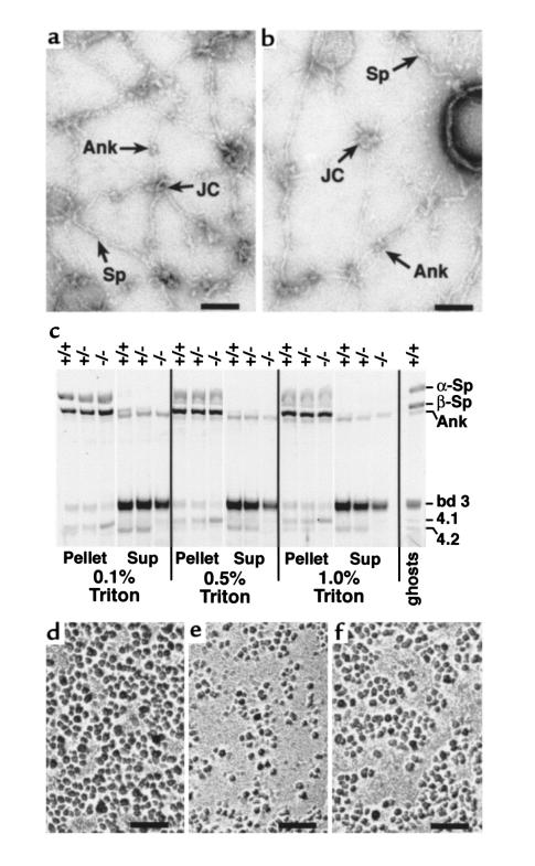
Figure 4.
Ultrastructure of RBC membrane skeletons. (a and b) Negatively stained, spread membrane skeletons from normal (a) and protein 4.2–/– (b) RBCs. Sp, spectrin filament. JC, junctional complexes. Ank, ankyrin. Scale bar: 100 nm. (c) Triton X-100–extracted RBC membranes. Ten micrograms of normal (+/+), heterozygous (+/–), and 4.2-null (–/–) RBC ghost membranes was extracted in 0.1, 0.5, or 1.0% Triton X-100. The pellet and supernatant (sup) fractions were resolved on a 3.5–17% Fairbanks gradient gel and stained with Coomassie blue. Sp, spectrin. Ank, ankyrin. bd 3, band 3. 4.1, protein 4.1. 4.2, protein 4.2. (d–f) IMPs in normal (d), band 3–null (e), and 4.2-null (f) RBCs. Note the presence of enlarged particle-free areas in band 3– and 4.2-null RBCs. Scale bar: 100 nm.

