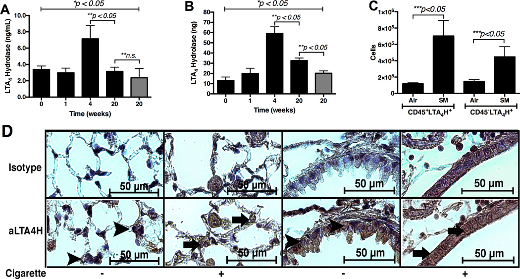
Figure 2.
Assessment of the LTA4H protein expression in 129sv wild type mice post exposure to cigarette smoke over 5 months. Panel A: The levels of LTA4H protein in the whole lung BALF assessed by ELISA. Panel B: The levels of LTA4H protein in the whole lung protein soup assessed by ELISA. Panel C: Flow cytometry of the whole lung single cell suspension. Air = mice exposed to ambient air for 20 weeks. SM = mice exposed to cigarette smoke for 20 weeks. Panel D: Immunohistochemistry with antibody specific to murine LTA4H protein in lung tissues from mice exposed to cigarette smoke for 20 weeks (63× oil magnification). Positive signals appear brown and counterstained with blue. Arrowheads show nuclei with positive counterstain (blue) indicating absence of LTA4H protein. Arrows show nuclei with positive stain (grown) indicating presence of LTA4H protein. Gray bars in Panels A & B = ambient-air exposed mice with ages 26 – 28 weeks. * represents analysis by ANOVA, ** by Bonferroni subgroup comparison, and *** by nonparametric t-Test. N = 5 per group.

