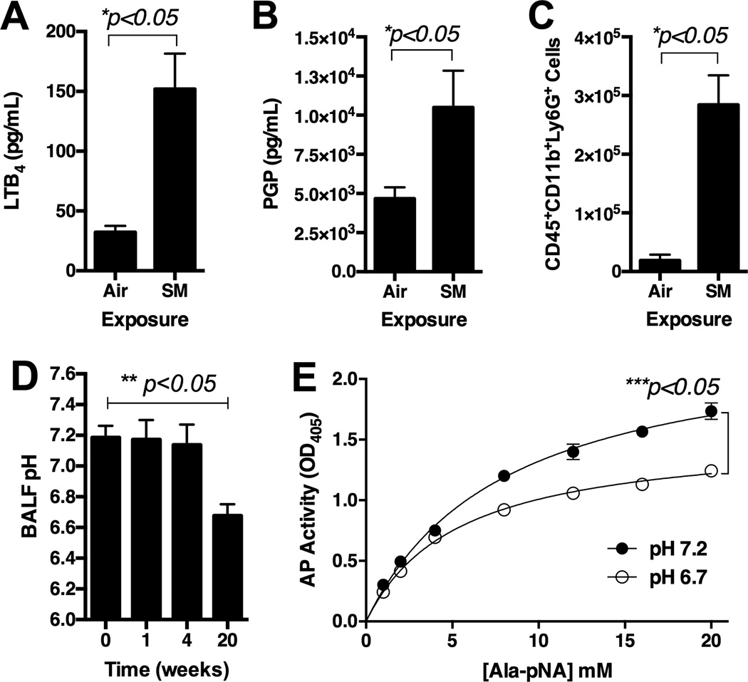
Figure 3.
Assessment of 129sv wild type mice post exposure to cigarette smoke by Teague TE-2 smoking apparatus for 5 months. Panel A: Levels of LTB4 in BALF. Panel B: Levels of PGP in BALF. Panel C: Levels of CD45+CD11b+Ly6G+ cells in whole lung single cell suspension. Antibodies are with PerCP-labeled CD45, ACP-labeled CD11b, and PE-labeled Ly5G. Panel D: pH of BALF over 20-week cigarette smoke exposure. Panel E: In vitro aminopeptidase activity assay using human recombinant LTA4H at pHs 6.7 and 7.2. * represents analysis by nonparametric t-Test. ** represents analysis by ANOVA.*** represents two-way ANOVA with AP activity and time as two factors. N = 5 per group. Air = ambient air exposure. SM = cigarette smoke exposure.

