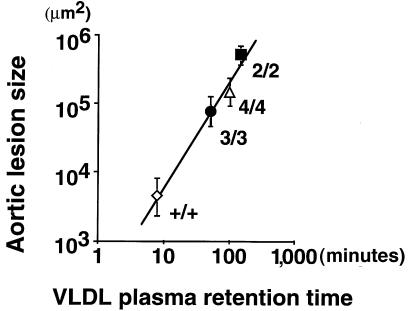
Figure 5.
Correlation between atherosclerotic lesion size and circulation time of VLDL particles. Atherosclerotic lesion area from at least 6 female mice maintained on an HFC diet for 3 months was determined. The log of the mean lesion size was plotted against β-VLDL retention time (inverse of the fractional catabolic rate; Figure 4 and refs. 15, 16), as determined from at least 3 mice injected with apo E–deficient labeled VLDL. The symbols are as follows: wild-type (open diamond), 2/2 (filled square), 3/3 (filled circle), and 4/4 (open triangle).

