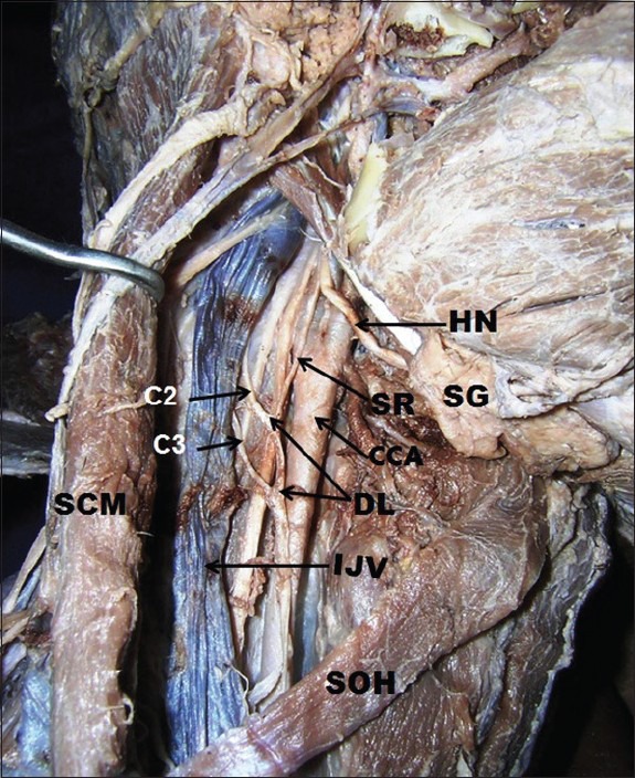
Figure 1.

Dissection of carotid triangle to show double looped ansa cervicalis associated with its deep topographical disposition beneath internal jugular vein. HN: Hypoglossal nerve, SG: Submandibular gland, CCA: Common carotid artery, SR: Superior root of ansa cervicalis, SCM: Sternocleidomastoid muscle (retracted backwards), SOH: Superior belly of omohyoid muscle, C2, C3: Second and third cervical nerves, respectively
