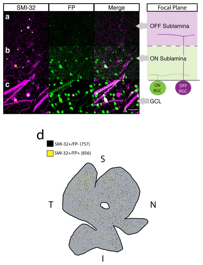
Fig. 1. A subset of SMI-32 immunopositive RGCs label with a melanopsin reporter.
(a–c) Retinas from adult Opn4Cre/+;Brainbow-1.0 mice in which all ipRGCs are labeled with FPs (Green) were immunostained for the alpha RGC marker SMI-32 (Magenta). Far right panels show the focal plane in which z-stacks in a–c were taken relative to the soma location or dendrite stratification of ON and OFF RGCs within the retina. (a) SMI-32+FP− cells (white *) stratified in the OFF sublamina of the IPL (white arrowheads). (b) SMI-32+FP+ cells (yellow *) stratified in the ON sublamina of the IPL (yellow arrowheads). (c) ipRGCs with the largest somas always colocalized with SMI-32 (yellow *), but not all SMI-32+ cells colocalized with FP (white *). (d) Wholemount Opn4Cre/+;Brainbow-1.0 retina immunostained for SMI-32. Both SMI-32+/FP+ (856, yellow dots) and SMI-32+/FP− (757, black dots) cells are found across all retinal quadrants (Coverage Factors: ON = 5.1 ± 1.3, n = 3 retinas, OFF = 4.9 ± 0.9, n = 3 retinas). Scale bar (a–c) = 50 μm. I = inferior retina, S = superior retina, T = temporal retina, N = nasal retina. GCL: ganglion cell layer, FP: fluorescent protein, RGC: retinal ganglion cell.

